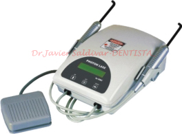
What is a laser and how does it work?
A laser is an instrument that produces a very narrow,
intense beam of light energy. When laser light comes in contact with
tissue, it causes a reaction. The light produced by the laser can remove
or shape tissue.
Are lasers used in dentistry?
Yes, lasers have been used in dentistry since 1990.
Lasers can be used as a safe and effective treatment for a wide range of
dental procedures and are often used in conjunction with other dental
instruments.
How are lasers used in dentistry?
Dental lasers can be used to:
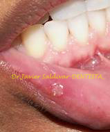
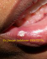
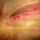
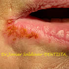
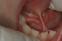
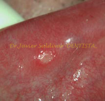
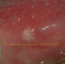
- reduce the
discomfort of canker and cold sores.
- expose partially erupted wisdom teeth.
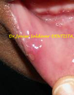
- remove muscle attachments that limit
proper movement.
- manage gum tissue during impressions
for crowns or other procedures.
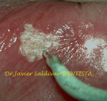
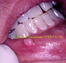
- remove overgrown tissues caused by
certain medications.
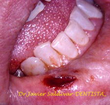
- perform biopsy procedures.
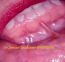
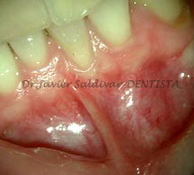
- remove inflamed gum tissues and aid
in the treatment of gum disease.
- remove or reshape gum and bone
tissues during crown lengthening procedures.
- help treat infections in root canals.
- speed up tooth whitening procedures.
What are the benefits of using dental lasers?
There are several advantages. Dentists may not need to
use a drill or administer anesthesia in some procedures, allowing the
patient to enjoy a more relaxed dental experience. Laser procedures can
be more precise. Also, lasers can reduce symptoms and healing times
associated with traditional therapies; reduce the amount of bacteria in
both diseased gum tissue and in tooth cavities; and control bleeding
during surgery.
Are dental lasers safe?
If the dental laser is used according to accepted
practices by a trained practitioner, then it is at least as safe as
other dental instruments. However, just as you wear sunglasses to
protect your eyes from prolonged exposure to the sun, when your dentist
performs a laser procedure, you will be asked to wear special eyeglasses
to protect your eyes from the laser.
How can I be sure my dentist is properly trained to use a
laser?
Ask your dentist questions about the extent of his or her
laser education and training. Make sure that your dentist has
participated in educational courses and received training by the
manufacturer. Many dental schools, dental associations, and the Academy
of Laser Dentistry (ALD) offer dental laser education. The ALD is the
profession's independent source for current dental laser education and
credentialing.
How will I know if treatment with a dental laser is an
option for me?
Ask your dentist. Although the laser is a very useful
dental instrument, it is not appropriate for every dental procedure.
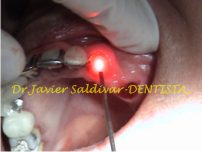
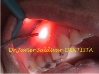
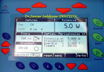
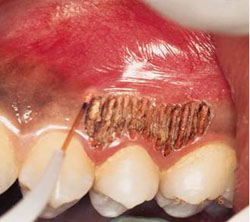
Virtually Painless, More Natural Dentistry*
Heat, vibration and pressure are the primary causes of
pain associated with the use of the traditional dental drill. Since
cutting both hard and soft tissues (teeth and gums) with LASER does not
generate heat, vibration or pressure, many dental procedures can be
performed nearly pain-free with fewer shots, less need for anesthesia,
less use of the drill and fewer numb lips! Additionally, using LASER for
gum procedures reduces bleeding, post-operative pain, swelling and the
need for pain medication in many cases. It’s just a better way to get
your dentistry done!
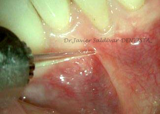
Accuracy & Precision
LASER dentists are able to remove tooth enamel decay (the
hardest substance in the body), bone and gum tissue precisely while
leaving surrounding areas unaffected. This conserves more of the
precious tooth structure you were born with, helping you to maintain
your natural teeth longer!
Reduced Trauma
High speed drills can cause hairline cracks and fractures
in the teeth that eventually lead to future dental problems. LASER
reduces damage to healthy portions of the tooth and minimizes trauma.
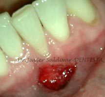
Less Bleeding & Swelling
Due to its conservative cutting action, LASER performs
many soft tissue (gum) procedures with little or no bleeding and less
post-op swelling.
Fewer Dental Visits
Since you often do not need shots or anesthesia, a LASER
dentist can perform cavity preps in all areas of the mouth in just one
visit. This technology also gives trained LASER dentists the ability to
perform many procedures that were previously referred to specialists,
saving you time and hassle as you address the dental needs of you and
your family.
Versatility
LASER is extremely versatile. It can be used for a wide
range of hard and soft tissue procedures. From decay removal, cavity
preparation, root canals, smile design, gum and bone surgical procedures
and many others.
*Discomfort is based on individual sensitivity to pain,
and other factors. Not all patients can be treated painlessly without
anesthetic. However, dentists using LASER to perform typical cavity
preparations report not using anesthetic in the majority of cases.
|


.jpg)
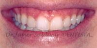
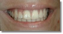
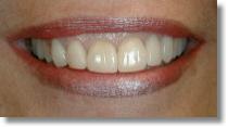
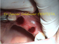
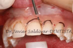
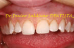
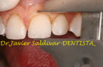
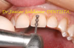
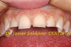
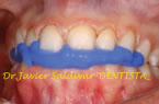
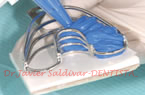
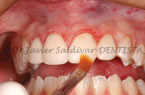
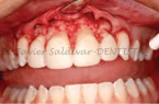
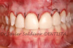
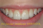
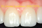
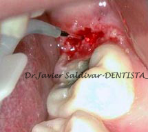
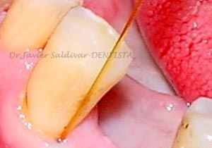
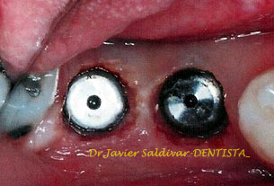
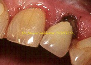
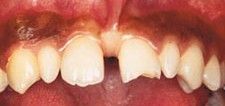
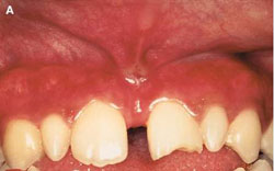
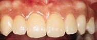
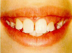
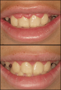
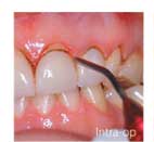
 The
picture here reveals how Dr.Saldivar skillful application of porcelain veneers
and use of Laser technology produces a near orthodontically correct smile.
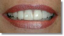
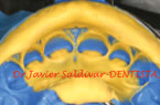
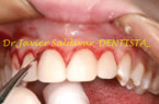
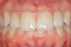
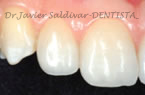
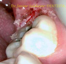
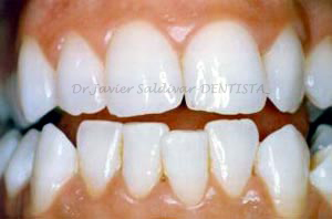
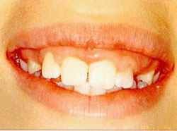
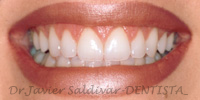
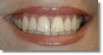
The
picture here reveals how Dr.Saldivar skillful application of porcelain veneers
and use of Laser technology produces a near orthodontically correct smile.